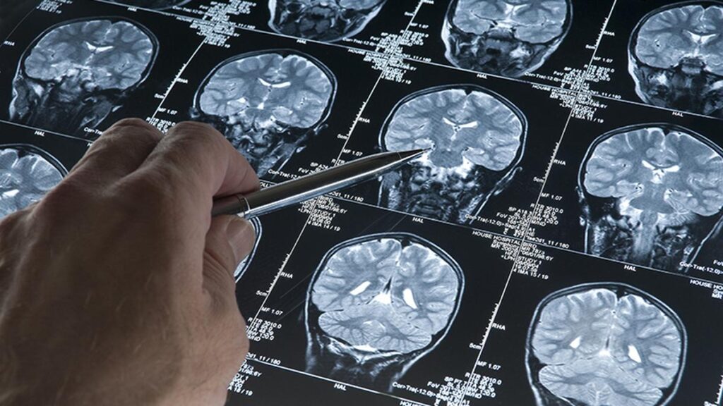
Timely MRI or CT scans can open the door to more treatment options and better outcomes for brain tumours. Photograph used for representational purposes only
| Photo Credit: Getty Images
A brain tumour is an abnormal mass or growth of cells in the brain. The sudden and unpredictable nature of brain tumours highlight how these conditions can affect individuals of any age, often without warning. These complex tumours range from benign growths to aggressive cancers. Increasing awareness about early symptoms and treatment options can dramatically improve clinical outcomes for patients, giving them a second chance at life.

Stories of hope
All patients with brain tumours have a unique story of how they faced their distinct set of challenges. Consider the story of Mr. G, a devoted father and businessman diagnosed with a right temporal glioblastoma. In an initial surgery, only the tumour’s core was removed, and the cancer margins regrew to their original size before radiation could be started. An MRI scan revealed that another surgery was needed. With an intraoperative ultrasound and neuronavigation, our team at was able to remove the tumour completely and safely. Today, Mr. G is stable and recovering well. He will soon receive Proton Beam Therapy — a form of targeted radiation.
Neuronavigation systems provide GPS-like precision inside the brain, while an intraoperative ultrasound helps locate the tumour in real time. Together, these tools have increased the extent of tumour removal — a factor closely tied to survival.

Some tumours hide in the brain’s deepest, most delicate regions. This was the case with 18-year-old K.S. from Ethiopia, who first underwent surgery and radiation for a brain tumour in 2019. When he returned to India in early 2024 with one-sided weakness, imaging revealed a cystic lesion with a calcified component deep within the midbrain. Pathology confirmed it as a benign pilocytic astrocytoma. After draining the cyst, there was some improvement but symptoms recurred a year later. Facing a recurrence in a high-risk location, an ultrasonic aspirator and meticulous microsurgical techniques were used to remove the lesion entirely. Now tumour-free, K.S. has returned home with a restored future. In hard-to-reach brain areas that control speech, movement, and vision, ultrasonic surgical aspirators are vital.
At 19, Mr. E experienced sudden seizures and foot paralysis. Scans revealed a 3 cm tumour situated in the right motor cortex, the area that controls voluntary movement. Preserving function in such delicate terrain is paramount. Surgeons used real-time neurophysiological monitoring with neuro-navigation and an intraoperative ultrasound to map critical brain regions and guide the resection. A tiny residual margin, deemed too risky for surgical removal, was scheduled for precision proton therapy. Neurophysiological monitoring tracks brain function during surgery, helping surgeons avoid damage to critical areas and preserve key abilities like movement and speech.

Modern advances in medical technology
Brain tumour care has evolved dramatically since the days of basic imaging and single-angle X-rays. Modern neuro-navigation systems function like an internal GPS, while intraoperative ultrasounds provide live visualisation of tumour boundaries. Ultrasonic aspirators rapidly break up tumour tissue with sound waves, enabling gentle suction and irrigation that shorten the duration of surgery.
In radiation therapy, proton beam treatment delivers radiation layer by layer, conforming tightly to the tumour, sparing surrounding tissues and reducing side effects by up to 60%, particularly in children. For patients who cannot undergo surgery or have deep-seated lesions, gyroscopic radiosurgery platforms offer fully non-invasive, outpatient treatments in sessions as brief as 30 minutes, targeting tumours from multiple angles with surgical precision.
No single specialty works in isolation. Each individual with a brain tumour diagnosis benefits from a multidisciplinary tumour board – neurosurgeons, medical and radiation oncologists, imaging specialists, pathologists, geneticists, and rehabilitation therapists collaborate on every diagnosis and treatment plan. Advanced technology and compassionate care go hand in hand, ensuring every patient feels informed and supported throughout their journey.

The power of early action
Brain tumours can be unpredictable: some remain stable for years, while others can double in size within weeks. Persistent headaches, sudden vision changes, seizures, memory lapses, or unexplained limb weakness should never be overlooked. Timely MRI or CT scans can open the door to more treatment options and better outcomes.
As cutting-edge technology continues to develop and care teams refine their expertise, the message is one of optimism that even in the most complex cases, there remains hope for healing. By turning early warnings into prompt action, patients can look forward to the brightest possible tomorrow with a chance at a new life!
(Dr. Ari G. Chacko is a senior consultant, neurosurgery and director of neurosciences, Apollo Proton Cancer Centre, Chennai. [email protected])
Published – June 21, 2025 04:00 pm IST

