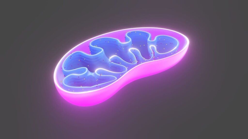
Mitochondria, the organelles in question, are best known as power packs—places where glucose molecules are disassembled to release the energy that drives metabolism. Boosting a failing cell’s metabolic processes by adding new mitochondria could thus be a smart move.
But that is just a start. These organelles, the descendants of bacteria that cosied up with the ancestors of human beings back when those ancestors were unicellular, retain from their days of independence a list of other jobs. These include disassembling surplus fatty acids and amino acids, and synthesising haeme, the active centre of haemoglobin and several other proteins.
Booster packs
Mitochondria also initiate the suicide of cells that are damaged, cancerous or surplus to requirements; act as communications centres for signalling proteins; and regulate levels of calcium ions—which are involved in signalling as well. They have their own genomes, too, separate from the main one in a cell’s nucleus. That is another legacy of their independent background.
With such a wide range of vital tasks to perform, it is hardly surprising that faulty mitochondria cause or contribute to many diseases. Some of these are congenital, the result of faulty mitochondrial genes. And some, such as diabetes and cardiovascular problems, occur when mitochondria wear out in old age. If a technique to transplant healthy ones could be made to work, its potential would be enormous.
One person trying to make this happen is James McCully of Harvard Medical School. He has developed a treatment for premature babies who, because the mitochondria in their heart muscles have been damaged by ischemia (the medical term for restricted blood flow), need the assistance of a heart-lung machine. Without such intervention, they would die. Even with it, only 60% survive.
In a trial, the results of which were published just over four years ago, Dr McCully improved that rate to 80%. His technique involves taking a small piece of tissue from the child’s abdominal wall, breaking it up to liberate the mitochondria, separating them from other cellular gubbins in a centrifuge and perfusing them back into the failing heart.
There is a chance that Dr McCully’s results may have been a statistical fluke—only ten babies were given the procedure in his experiments—but it suggests his technique is at least safe. He and his colleagues found that their procedure immediately increased production of signalling molecules in the babies, which stopped inflammation and cellular suicide. And, shortly afterwards, the perfused mitochondria took up residence in the damaged heart muscle, restoring its function in the longer term.
Recharge and refresh
Dr MCully now hopes to extend this approach, which is currently being assessed by America’s Food and Drug Administration, to other ischemia-affected tissues, including adult hearts, lungs, kidneys and limbs. Nor is he alone. Lance Becker of the Feinstein Institute in New York plans to test a similar technique on premature babies. And Melanie Walker of the University of Washington, in Seattle, has just published the results of an experiment on a different type of ischemia—that which causes strokes.
This trial, reported in November 2024, was conducted mainly to check safety (in which regard it passed), so it involved only treating four participants. But Dr Walker says early indicators of efficacy were “promising”. Her technique involved infusing the site of the ischemia-inducing blood clot with mitochondria as part of an otherwise-standard procedure to remove the clot. The intention, which she hopes to test in a future trial, is to stop neurons affected by the stroke from killing themselves.
Dr Walker has further trials on the slipway. One is for adult hearts. Another aims to restore function to neurons injured by physical trauma rather than strokes. And a third is for Pearson’s syndrome, a congenital combination of anaemia and pancreatic problems caused not by trauma but rather by the deletion of a stretch of DNA from the mitochondria of those who have it.
Such mutations are rare. Normally, a mother’s mitochondria are passed intact to her offspring via her egg cells. Sometimes, however, a mutation occurs spontaneously on the way to an egg’s creation, meaning the resulting offspring may have symptoms that their mother does not. Dr Walker plans to pick patients with unaffected mothers and enrich blood-forming stem cells taken from those patients with mitochondria extracted from their mothers’ white blood cells. The enriched cells will then be returned to the patient, where they will, with luck, give rise to healthy blood cells that relieve the anaemia.
Congenital deletion-related conditions such as Pearson’s affect about one person in 5,000. That is a number big enough to interest aspiring biotech firms. Minovia Therapeutics, an Israeli company, has Pearson’s in its sights, along with Kearn-Sayre syndrome (KSS), another deletion-related condition, and myelodysplasia, a form of anaemia caused by mitochondrial mutations that happen later in life.
Preliminary trials using the method Dr Walker plans to adopt relieved symptoms of Pearson’s and KSS in children. A new approach, in which the mitochondria are extracted from discarded placental tissue rather than from living human beings, is now being tested for myelodysplasia.
Those involved in these projects hope that, besides relieving anaemia, the reinvigorated stem cells may also pass their mitochondrial cargo on to other affected tissues. This is a hope based on the knowledge that such transfers occur naturally during the formation of blood cells.
Indeed, they also occur during wound healing, the creation of new blood vessels and the boosting of heart muscle. It thus seems plausible that the body contains a sophisticated, hitherto unperceived, mitochondrion-transfer network, in which some cells act as mitochondrion nurseries, releasing their products into the bloodstream for the benefit of cells that cannot generate enough of them internally. Certainly, blood contains huge numbers of free-floating mitochondria—one study suggested perhaps as many as 3.7m per millilitre.
At an earlier stage of development than the human trials, meanwhile, are a range of promising experiments using cell cultures and laboratory animals. Aybuke Celik, a colleague of Dr McCully at Harvard, is investigating the effect of transplanted mitochondria on prostate- and ovarian-cancer cells. She has found they reduce the amount of chemotherapy needed for such cells to kill themselves.
Conversely, a team at Zhejiang University in Hangzhou, China, used rats to show that transplanted mitochondria stop damaged neurons pressing the self-destruct button—an observation that may one day help people with spinal injuries avoid paralysis.
Longing for longevity
One of the most intriguing findings of all, though, is that—in laboratory cultures, at least—transplanted mitochondria rejuvenate the biochemistry of elderly host cells. Given the number of free mitochondria in blood, this may help explain the puzzling observation that transfusing blood plasma from young to old animals seems to grant the latter a new lease of life.
This observation has long excited people seeking to prolong human “healthspan” to match the extended lifespans now enjoyed in rich countries. But the search for the elixir involved has hitherto focused on the plasma’s molecular cargo. Perhaps it is not molecules but mitochondria that would-be Methuselahs should consider.
© 2025, The Economist Newspaper Ltd. All rights reserved. From The Economist, published under licence. The original content can be found on www.economist.com

