
In a new study, scientists at the Indian Institute of Technology (IIT) Bombay have demonstrated how cells can sense and respond to invisible mechanical patterns—like built-in tensions around them.
The research led by Professor Abhijit Majumder, was published in Cell Reports Physical Science. The findings not only add to the fundamental understanding of how cells organise themselves, but also have important implications for tissue engineering, cancer research, and wound healing.
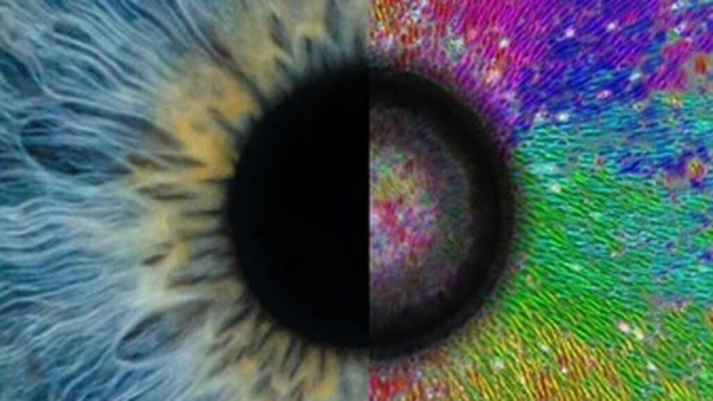
Cells follow very specific patterns, for instance, muscle fibres are aligned parallel to each other to enable coordinated movements, blood vessels extend toward wounds to facilitate healing, and cells in the eye are arranged radially to help focus light precisely onto the retina, ensuring clear and accurate vision. Such precise spatial organisation is essential for proper tissue function.
The arrangement of cells directly influences how effectively a tissue can carry out its role, be it contracting, transporting nutrients, or processing sensory input. But how do cells determine their correct location and orientation within these complex systems? Professor Majumder said that for decades, scientists believed that cells primarily relied on chemical signals, like growth factors or morphogens, to decide how and in which direction to grow.
“However, recent discoveries in this field suggest that mechanical signals are just as important. Cells can feel how stiff their surroundings are, detect tiny stretches, and even respond to surface textures smaller than themselves. In living tissue, mechanical inhomogeneities are common. You see it in tumours, healing wounds, and developing organs. But we haven’t fully explored how cells interpret and respond to these physical cues,” Professor Majumder said.
The researchers embedded a rigid object inside an otherwise soft material, mimicking mechanical inhomogeneity. The goal was to mimic how tissues naturally develop internal tension during processes like growth, injury, or tumour formation, and how cells might sense and respond to such forces.
Lead author Dr. Akshada Khadpekar explained, “To simulate these conditions, we designed a soft polyacrylamide hydrogel with a small, rigid glass bead embedded inside. This setup replicates a rigid structure surrounded by softer material, like a tumour within the body tissue.”
When the gel was placed in water, it began to swell everywhere except where the bead was, because the stiff bead resisted the expansion. This created a pre-strain gradient— a varying stretch pattern around the bead.
When muscle precursor cells were added to the gel, the pre-strain gradient played a crucial role in guiding their alignment.
“Cells near the bead detected the pre-strain gradient and aligned radially. As they exerted forces on the substrate, the mechanical signal propagated outward. This alignment extends about 1–2 mm (20–40 cell lengths) from the bead, reinforcing long-range organisation,” Dr. Khadpekar said.
On a soft, uniform gel without a bead, however, the alignment was limited to about 0.35 mm. Professor Majumder added, “Think of it like a shallow crater around the bead. But instead of falling in, the cells sense the stretching pre-strain and align accordingly.”
To be sure this wasn’t due to chemical factors, the researchers ran control experiments. They changed the type of extracellular matrix (ECM) proteins used to coat the gel and varied the stiffness of the substrate. Only on soft gels did the cells align. Harder gels masked the effect, and altering the ECM had no impact, proving that the alignment was not biochemical in origin.
To further investigate the mechanism behind cell alignment, the research team collaborated with Professor Parag Tandaiya from the Department of Mechanical Engineering at IIT Bombay and employed finite element simulations to model the mechanical environment created by the swelling hydrogel. These computational models confirmed that the strain fields generated in the gel closely matched the patterns of cell alignment observed in experimental conditions.
“This was crucial because there’s no direct way to measure these subtle internal pre-strain fields experimentally. Without simulations, we wouldn’t have been able to formulate or test our hypothesis about what the cells were sensing,” Professor Tandaiya said.
To test the generality of this phenomenon, the researchers extended their experiments beyond individual spherical beads to hollow glass capillaries, glass beads, and their combinations.
In all cases, the cells aligned along invisible force patterns, forming arcs, waves, or spirals. The researchers also tested different types of cells and found that how the cells aligned depended on how much force they could apply and how stretched out they were.
“The cells don’t just sense stretching of their substrate, they seem to also detect the direction in which the substrate is stretched the most and they line up in that direction. It’s a very precise and intelligent response, and we believe this is the first time such behavior has been observed in this way,” Professor Tandaiya added.
Using these findings, a model was created to predict which cells would align based on their shape, strength, and stiffness of the surface. This discovery has significant implications across multiple fields.
“In tissue engineering, we might guide cell organisation just by shaping soft materials, without complex scaffolds or stimulation. Or in cancer, the stiffness of tumours could explain how they influence nearby cells. And in regenerative medicine, adjusting tissue stiffness may help restore healthy cell patterns in aging or damaged tissues,” Dr. Khadpekar said.
Published – June 11, 2025 08:00 am IST